
| Incredible Horizons: New Brain Research | ||||||
| Brain Plasticity- The good news is that: The brain is can repair itself through specific appropriate input through the senses. If you feel like you have "faulty wiring", we can help your brain function more efficiently. Scientists apply the term neuro-plasticity or brain plasticity to the action of brain growth and adaptation Our products produce better grades with less effort for every subject the rest of their life
| ||||||
|
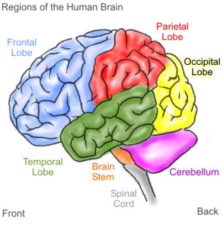
| Science has repeatedly demonstrated that the brain can change and grow given the right learning tools and environment. Our programs are founded in educational cognitive psychology to provide the correct environment and challenge.Get Attention strives to provide the very best learning tools for the creation As recently as twenty years ago, scientists believed that the genes we were born with wholly determined the structure of our brains. However, current extensive research performed by scientists worldwide proves that how our brains develop, learn, and grow depends on the vital interaction between nature Neural Networks Learning takes place by construction of new neural networks/pathways. Neural networks are the “whispering” of neurons to each other. Neurons are brain cells that communicate with each other Brain related research continuously strives to deepen our understanding of brain functions. We have learned that new information (sensory input) enters the brain through preexisting networks, which is why it is imperative to provide challenging stimulation in early childhood. If the input is not new, it can trigger memory. If it is new, it can trigger learning thus creating new pathways. Cognitive psychology refers to this process as constructivism: The learner builds his or her own knowledge on his current knowledge base, but only in response to a challenge and/or new stimulation. It is evident that some persons are not born with the neural networks that facilitate focused attention. Our cognitive training, light and sound and neurofeedback programs are designed to directly challenge users to build new neural networks necessary for optimum performance.
Neural networks challenged/stimulated through our light and sound equipment, can enhance physical healing and homeostasis while increasing attentional flexibility. As early as the 1930’s, there was research showing that repetitive pulsing lights and percussive sounds will stimulate and synchronize the hemispheres of the brain. This creates a frequency following response known as entrainment. Through flickering lights and controlled rhythmic tones, we can entrain the brainwave frequencies into desired states of consciousness. With the relief of tension or resistance in the mind/body, the user becomes receptive to information and the ability to process and recall it. Once the balancing and stress reducing effects of the equipment become engrained, a user can then proceed to special applications Link to our home programs that help rebuild the brain 12/03-Update-New Research has found physical markers in biochemical balance Our neuro-supplement Balance Formula 1 is the perfect supplement to help with this. Our entire Dr. Allerton recognized that in order for the brain to function properly and for the neurotransmitters If you are at a stand still or plateau with other therapies-ie. ABA, Floortime etc.-You have probably benefited all you can from that therapy until you improve how the brain actually functions. Programs Brain state research for the attentionally challenged Every thought, feeling, sensation, and level of awareness has a corresponding brain wave pattern. There are brainwaves that are considered slow brainwaves such as Delta, Theta, and Alpha and fast brainwaves’ like SMR and Beta. Here is a chart of to help explain brainwaves. Frequency of Mental States Description of Brainwave States Brainwaves DELTA: Sleep The ‘Sleep’ State 0.5-3(cps)cycles per second THETA: Inner reflection w/o much atten- The ‘Tuned-Out’ 4-7 (cps) tion focused on the outside world; Waves tuned out; drowsy ALPHA: Resting in a meditative & creative The ‘Daydreaming’ 8-11 (cps) state; daydreaming; inattentive Waves SMR: Calm, not fidgeting, not impulsive, The ‘Calm’ Waves 12-15 (cps) not thinking about bodily sensation; Often externally aware; quietly alert BETA: Focused analytical, often externally The ‘Thinker’ Waves 16-20 (cps) oriented, intense thinking/processing Slow waves indicate daydreaming and fast waves indicate concentration in a normal person. When we are inattentive or daydreaming, our brains produce primarily slower brainwaves like theta and alpha. As we focus on a task like reading or listening, our brain engages and these slow waves ‘drop out’ with brain energy moving to the faster ‘thinker’ waves. Consequently, our brain ‘wakes up’ and becomes activated to process this information. The ADHD brain works differently. Research demonstrates that when an ADHD child tries to concentrate, the brain continues to produce even more slow wave activity leading to a state of under-activation or low arousal. Have you ever wondered why ADHD children can focus so intently on video games, TV, or things they are interested in, yet cannot focus on their schoolwork? I have. There is a theory called the low arousal theory. The ADHD individual produces excessive levels of Theta or “Slow Brainwave” patterns that act like a fog or filter. High levels of stimulation/physical activity penetrate through the fog activating attention. As a child is engaged in a stimulating activity, like playing a video game, there is enough stimulation present in the activity to penetrate through the slow wave ‘filter’ to activate arousal-directing attention. During a ‘low’ stimulation task like reading, doing homework or chores, there is not as much stimulation coming from the environment. Now there is not enough stimulation present in the activity to penetrate through the slow wave ‘filter’ to activate arousal, so it is almost impossible to direct attention. A low level of stimulation results in boredom. The boredom leads to increased activity. Children with ADHD are dependent on stimulation from the environment to direct attention – they are unable to self-regulate. If there is not sufficient stimulation present from the environment, these children seeks out or create stimulation around them. The child may become fidgety, restless, get out of their seat, talk to their neighbor…these activities are designed to increase stimulation to break out of this state of low arousal. Neurofeedback can help to actually assist an individual in changing, (controlling) physiological differences found in those suffering from ADD/ADHD. Everyone experiences this cycle to some degree. Can you recall an experience of eating lunch or dinner, then having to sit in a classroom or attend a meeting? As your metabolism begins to kick in, you get a bit drowsy and begin to daydream. You are beginning to produce more alpha brainwaves. Now as you are forced to sit there, the metabolism kicks in, your eyes become heavy, you are feeling so drowsy it is hard not to fall asleep. You are now producing more theta brainwaves. Because you are so drowsy and drifty, you are not as likely to receive and assimilate this information, as well as when you are in a more alert and wide awake state. You probably want to get up, move around, get a cup of coffee, – and do something to wake up! This is what happens to the ADHD child. As he sits there in a low stimulation environment, like a classroom, or doing homework or chores, the brain continues to produce slower brainwave activity. Vanderbilt study could lead to understanding behavior For more info on healthy- drug free programs for successful living click on a link below Incredible Horizons Articles Loop |
<>+<>+<>+<>+<>+<>+<>+<>+<>+<>
~The Cosmic-Visions Blog~


 Encyclopædia Britannica Article